
Taking advantage of the mechanical differences between healthy tissue and tumors, a wireless probe can aid surgeons in precise tumor removal in breast cancer.
From the Journal: APL Bioengineering
WASHINGTON, May 5, 2026 — Breast cancer impacts over 2 million women around the world each year. Following radiotherapy or chemotherapy, breast-conserving surgery is the first line of intervention for early-stage breast cancer. This surgery aims to remove the tumor while preserving as much of the healthy tissue as possible, but since precise tumor mapping during surgery is challenging, sometimes the tumor is not fully extracted.
Without proper removal, patients face future surgeries, delayed follow-up treatments, and a decreased quality of life.
In APL Bioengineering, by AIP Publishing, researchers from the University of Western Australia, the University of Melbourne, the Royal Melbourne Hospital, and Nicolaus Copernicus University in Toruń designed and tested a hand-held device to differentiate tumors from healthy tissue using the tissues’ mechanical properties.
Often, tumors are stiffer than benign tissue — a difference that can be felt during surgery.
“The technique presented in this paper — stereoscopic optical palpation (SOP) — was inspired by clinical palpation, which is routinely used by surgeons to identify diseased tissue based on the sense of touch,” author Rhys Jones said.
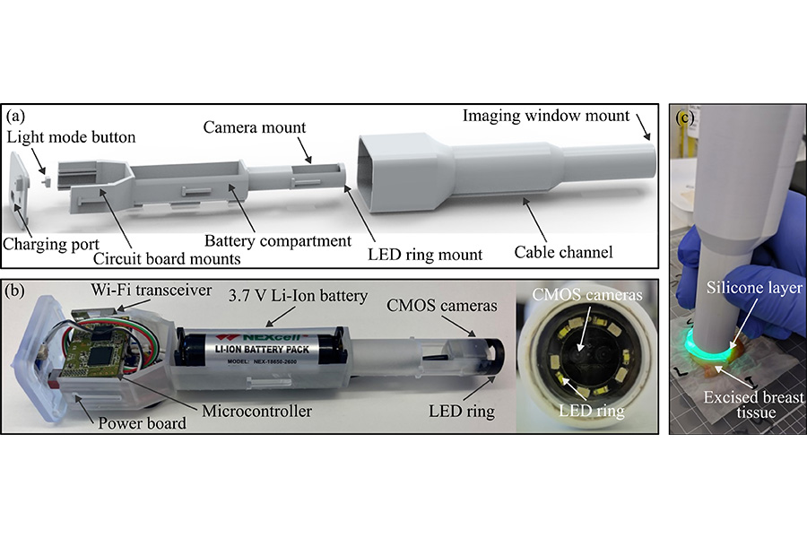
The hand-held probe the researchers developed is based on optical elastography, a field that combines imaging with mechanical measurements like elasticity. When compressed, tumors and healthy tissue exhibit different mechanical properties, and this can be measured by the probe. Since the probe also provides a visual of the tissue, users have another data point to differentiate the two tissue types.
“The wireless probe is an extension of previous benchtop implementations of SOP,” Jones said. “Our process began with establishing key requirements for the probe through consultation with surgeons: We decided that our targets were an ergonomic hand-held format, a field of view of at least 6 millimeters by 6 millimeters, wireless operation, a minimum one-hour battery life, and low material costs.”
The material cost of the prototype was around $1,200, which is less than similar benchtop SOP devices priced around $3,000, but the authors believe this can be reduced if it is mass-produced. Ultimately, Jones wants the device to be used in vivo during surgery, even going beyond tumor removal.
“Beyond breast-conserving surgery, we believe that the probe could be used in many clinical scenarios where palpation is currently used, including the assessment of skin lesions,” Jones said.
###
Article Title
A wireless and handheld optical palpation imaging probe for use in breast-conserving surgery
Authors
Rhys Jones, Renate Zilkens, Ankit Bharakhda, Mireille Hardie, Christobel M. Saunders, Qi Fang, Brendan F. Kennedy
Author Affiliations
University of Western Australia, University of Melbourne, Royal Melbourne Hospital, Nicolaus Copernicus University in Toruń