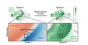
Visualizing electrical patterns underlying abnormal heart contractions and deformations.
From the Journal: Chaos
WASHINGTON, D.C., September 17, 2019 — Despite advances in medical imaging, the mechanisms leading to the irregular contractions of the heart during heart rhythm disorders remain poorly understood.
Research from the University of Göttingen in Germany suggests existing data from ultrasound imaging can be used to work backwards to reconstruct the underlying electrical causes of arrhythmias. The scientists describe the work in Chaos, from AIP Publishing.
“The heart wall is pretty thick, and current imaging can’t see through it,” said author Jan Christoph, a biomedical physicist at the University Medical Center Göttingen. “Cardiologists can only map the surface of the heart by inserting catheters into a patient’s heart, and it’s currently impossible to measure the electric activity deep within and throughout the heart muscle all at once.”

However, to better locate possible origins of heart rhythm disorders, cardiologists need to be able to look deeper into the tissue. To help overcome this obstacle, Christoph and his colleagues tested a computational approach to see if it would be possible to extract information about the electrical activity within the heart without directly observing it but inferring it from the heart’s mechanical deformations. They believe the approach could be combined with ultrasound or MRI imaging.
The researchers made computer models of two systems interacting with each other – one representing a piece of a heart wall with a rhythm disorder and the other representing a virtual heart. In the virtual heart, the electrical activity is carefully adjusted so it starts to deform in the same way as the arrhythmic heart. Similar approaches have been used in computer weather predictions, in which measurement data from weather stations is assimilated by computer models.
By studying the mechanical characteristics of the two heart models, the researchers found they were able to reconstruct the initial electrical wave patterns almost exactly in the arrhythmic heart.
Applied to a heart, this means studying mechanical cardiac behavior — such as the contractions and deformations that characterize arrhythmias — may reveal information about the electrical activity within the heart previously out of reach. “That would give you the hidden electric excitation,” said Christoph.
Though the work has only been tested in computer simulations so far, the researchers anticipate the reconstruction approach can soon be applied when imaging patients. The group will be conducting a preliminary study on the feasibility of the method on patients who suffer from heart rhythm disorders, such as ventricular tachycardia, with medical colleagues in Göttingen and Hamburg.
“We want to image patients as they undergo ablation procedures using 3D ultrasound imaging,” Christoph said. “We then hope we can apply the reconstruction technique to estimate the abnormal electrical patterns throughout the heart.”
For more information:
Larry Frum
media@aip.org
301-209-3090
Article Title
Synchronization-based reconstruction of electromechanical wave dynamics in elastic excitable media
Authors
Jan Lebert, Jan Christoph
Author Affiliations
University Medical Center Göttingen
