From the Journal: APL Bioengineering
WASHINGTON, August 3, 2021 — Ovarian cancer devastates more than 20,000 women in the U.S. every year, due in part to its tendency to evade detection and present after metastatic spread. A key element to slowing metastasis is understanding the mechanisms of how tumor cells invade tissues.
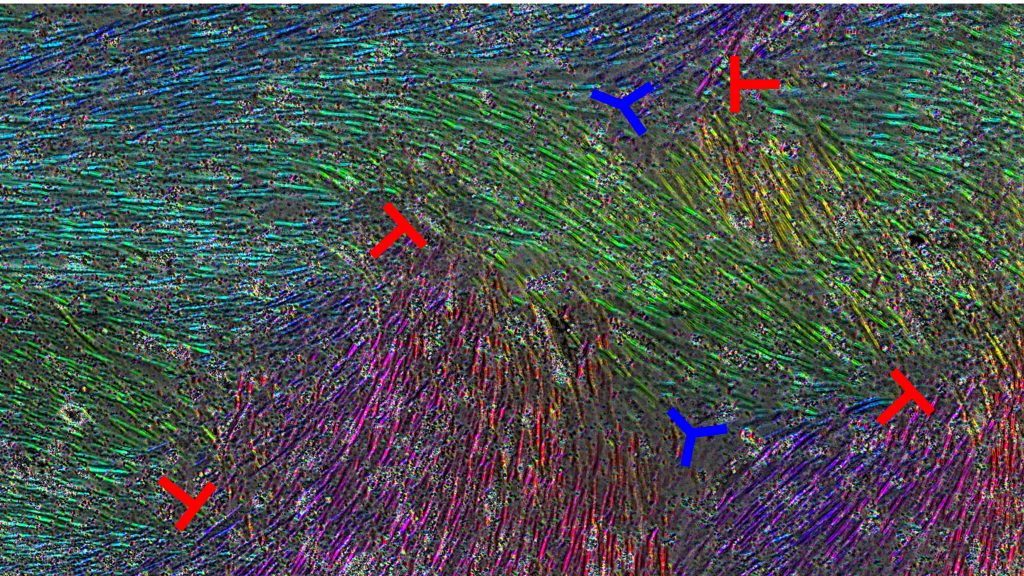
In APL Bioengineering, by AIP Publishing, biophysics researchers at the University of Wisconsin explain how microscopic defects in how healthy cells line up can alter how easily ovarian cancer cells invade tissue. Using an experimental model, where the cellular makeup mimics the lining of the abdominal cavity, the group found that disruptions in the normal cellular layout, called topological defects, affect the rate of tumor cell invasion.

“My lab is very interested in identifying ways to slow metastasis. This study is exciting, because it demonstrates a unique role for organization of nontumor cells to either aid or slow that process,” said author Pamela Kreeger. “Identifying factors that regulate this organization could help us to achieve our goal.”
Topological defects are well known to the world of physics, ranging from quantum field theory to cosmological phenomena, but are only starting to find use in medicine and biology.
The group’s model consisted of a single layer of healthy cells, called mesothelial cells, the predominant cell type that covers structures inside the abdomen, where ovarian cancer often metastasizes.
“A common way to fill space is a honeycomblike packing, in which each ‘cell’ would be nearly spherical,” said author Jacob Notbohm. “But in our case, the mesothelial cells were elongated, making the honeycomb packing not possible.”
Such elongation led to areas of well-ordered cell layers and left other areas with alignment imperfections, causing the topological defects. These flaws in this alignment have been associated with a host of microscopic influences, including altered cell density, motion, and forces.
They seeded ovarian cancer cells on top of the mesothelial cell layer and compared what effect the arrangement of the mesothelial cells had on how the tumor cells passed through this barrier.
The patterns of cell flow were different near the defects, with certain defects causing inward cell flow, toward the center of the defect. At those locations of inward flow, the cancer cells passed through the mesothelial barrier more slowly.
In addition to pursuing the impact of topographical organization in cancer cell metastasis, the group is looking to investigate the cause of topological defects, with the hopes of finding ways to direct cell patterning in uses, such as tissue engineering.
###
For more information:
Larry Frum
media@aip.org
301-209-3090
Article Title
Topological defects in the mesothelium suppress ovarian cancer cell clearance
Authors
Jun Zhang, Ning Yang, Pamela Kreeger, and Jacob Notbohm
Author Affiliations
University of Wisconsin
