From the Journal: APL Bioengineering
WASHINGTON, June 2, 2020 — Advanced tissue engineering technologies allow scientists to mimic the structure of a uterus, enabling crucial research on fertility and disease.
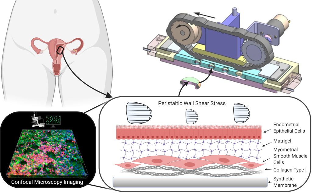
In an APL Bioengineering article, by AIP Publishing, researchers present two mechanobiology tools for experiments on synthetic or artificial uterine tissue. They wanted to study the negative effects of hyperperistalsis, contractions of the uterine wall that occur too frequently.

Throughout an individual’s lifetime, the uterus undergoes spontaneous contractions of the uterine wall, which can induce uterine peristalsis, a specific wavelike contraction pattern. These contractions are important for many reproductive processes, such as the transportation of sperm prior to impregnation, but hyperperistalsis could impede fertility and lead to diseases, such as adenomyosis or endometriosis.
“The nonpregnant spontaneous myometrial contractions induce the uterine peristalsis, which exerts physical loads on the internal endometrial barrier and, thereby, affect the biological performance,” said David Elad, one of the paper’s authors.
The designed tools include a well, which can be disassembled for installation of a biological in vitro model in an experimental chamber, and a flow chamber and transmission system that create the contraction patterns.
“The cells are cultured on a biological or synthetic membrane stretched over a well bottom, which is installed into a cylindrical medium holder. Once the biological model is ready, the well bottom can be disassembled and then installed in the experimental chamber,” said Elad. “As far as we know, it is the first time an in vitro biological model was exposed to peristaltic wall shear stresses.”
Using their experimental setup, the authors found peristaltic shear stresses caused alterations to the cytoskeleton of endometrial epithelial cells and myometrial smooth muscle cells of the synthetic uterine tissue.
Future research using this approach might include studying the effect of different contraction patterns and the role hormones play in uterine peristalsis. The researchers noted uterine wall models can typically be applied to study models of intestine walls and some organs and tissues.
“The laboratory approach of this work demonstrates future options to explore the complex processes of human reproduction, especially during early stages when accessibility and ethical limitations prohibit in vivo experiments,” said Elad.
###
For more information:
Larry Frum
media@aip.org
301-209-3090
Article Title
Tissue engineered endometrial barrier exposed to peristaltic flow shear stresses
Authors
David Elad, Uri Zaretsky, Tatyana Kuperman, Mark Gavriel, Mian Long, Ariel Jaffa, Dan Grisaru
Author Affiliations
Tel Aviv University, Institute of Mechanics at the Chinese Academy of Sciences
